SHARES

Some people are born healthy, some are born with congenital defects, like a hole in the heart. It is commonly heard that many people had this condition when they were young, and some still have it while they age. The question is, where exactly is the hole? Will it heal by itself? And does this affect the daily lifestyle? This condition is called ventricular septal defects (VSDs). It is necessary to understand the structure of the heart to truly figure out this condition.
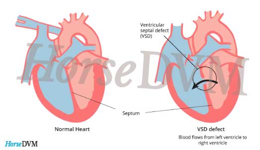
Where is it?
The heart has four chambers, two atria (upper chambers) and two ventricles (lower chambers). The atria function to receive blood and the two ventricles to pump blood out from the heart. The right ventricle pumps to the lung for gaseous exchange while the left ventricle supplies to the rest of the heart. The two ventricles are thick-walled chambers and during pregnancy the wall did not fully develop and thus forming a hole in between. In fact, the hole can happen in any wall of the heart but in this case, which is more common, it is known as ventricular septal defect (septal also means partition). This is a congenital heart defect. Congenital means present at birth.

Will it close by itself?
There are four possibilities, as follows:
- The hole may close spontaneously. This occurs in perhaps as many as 50 per cent of cases, usually in early childhood but sometimes in later childhood, adolescence or adult life.
- The hole may retain the same size. Since the heart is growing throughout childhood, the defect becomes relatively small.
- Due to the difference of pressure on both sides of the ventricles (left ventricle has higher pressure to pump blood for the rest of the body), eventually the pressure will reverse and the child may not receive enough oxygenated blood and may become cyanosed (bluish discoloration of skin and mucous membrane due to lack of oxygen in the blood).

- In long term, pulmonary hypertension will occur. It is defined as the increase in blood pressure that affects the arteries in the lung and right side of the heart. With this, the child will develop fainting episodes, shortness of breath, chest pain and so on.
Investigations:
– Chest X-ray: Produces image of chest.
– Electrocardiogram (ECG): Records the electrical activity of the heart.
– Echocardiogram: Ultrasound of the heart to show structure of the heart.
Types of Ventricular Septal Defect
Small VSD: The child will not develop any symptom since the shunt is small. However, there is a risk of getting infected over the valve of the heart (google bacterial endocarditis) and therefore preventive antibiotic is necessary at the time of dental extractions.
Moderate VSD: Symptoms usually occur in infancy, the breathlessness on feeding and crying, recurrent chest infections. As the child gets older, the symptoms tend to improve and may disappear altogether due to closure of the defect.
Large VSD: Affected babies might be severely ill and may develop heart failure. Surgical closure is sometimes needed. Surgery is usually performed at 3-6 months of age in order to manage heart failure and prevent permanent lung damage from pulmonary hypertension. After surgery, the doctor will set up regular follow-up visits to make sure the hole remains closed and symptoms remain minimal.
What to watch out in the future?
Apart from regular follow ups, there is no need to restrict activities unless warned by the cardiologist. These days, having a VSD is usually nothing to worry about. The cardiologist is who you can get all your questions answered from. Most people who have had a VSD can enjoy doing the same activities like others so it need not be a reason for people to hide under the roof and “do nothing but rest”.
Reference:
by Angie Loh
A medical student with nothing but passion and a pen. Poems and novels never fail to make me feel alive. I'm inspired to make the world a better place and fill it with a little bit more love. But first, where's my coffee? View all articles by Angie Loh.


